Research facilities
- Hyper-Raman spectroscopic system
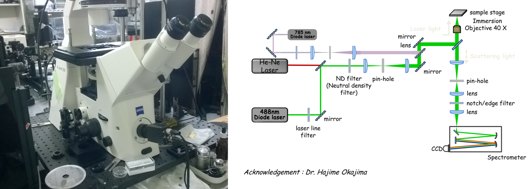
- Multi-wavelength Confocal Raman micro-spectrometer
- Confocal Raman microspectrometer (632.8 nm)
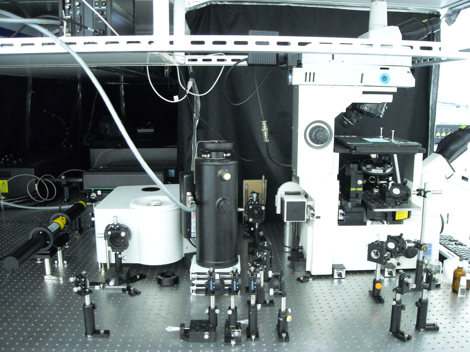
This apparatus is used to perform space-resolved Raman spectroscopy with sub-µm lateral resolution and a few µm axial resolution. The system consists of a cw He-Ne laser, a custom-made microscope, an imaging spectrometer, and a liquid-N2 cooled CCD detector. We collaborated with Nikon Instech to design the microscope. It combines the upright part with a normal, inverted microscope so that forward detection as well as backward detection is possible. We can measure Raman spectra at distinct locations on a living cell and generate a “Raman image” of the cell by means of a piezoelectric nanopositioning stage.
Reference: C.-K. Huang, H. Hamaguchi, and S. Shigeto, Chem. Commun. 47, 9423 (2011); Noothalapati Venkata, H. N., Nomura, N. and Shigeto, S. , J. Raman Spectrosc., 42, 1913 (2011)
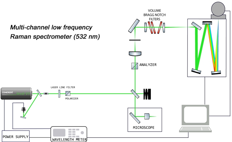
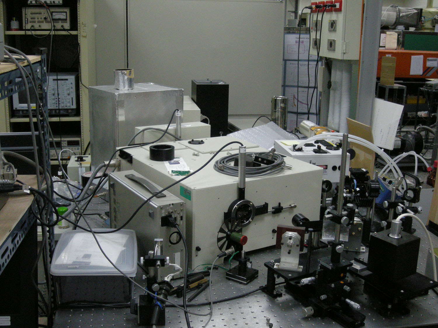
- Multichannel low-frequency Raman spectrometer
Raman spectra in the low-frequency region (<200 cm−1) provide a wealth of information on intermolecular vibrations and lattice vibrations of molecular and ionic compounds in the condensed phase. Real-time tracing of the low-frequency Raman spectra is therefore a powerful tool for understanding phase transitions such as melting and freezing from a molecular viewpoint. However, observation of low-frequency Raman spectra is severely hindered by immense Rayleigh scattering. In addition, to be able to keep track of dynamic phase transition processes, fast multichannel detection rather than single-channel detection with a double/triple monochromator is required. A low frequency system using volume bragg notch filters was developed by Dr. Hajime Okajima. Using this spectrometer the low frequency bands of L-cystine upto +/- 9.8 cm-1 can be observed. This spectrometer is also used for phase transition studies in polymers as well as rotational Raman spectroscopy of gases including nitrogen, hydrogen and more.
References: Okajima, H., and Hamaguchi, H.-o. , J. Raman Spectrosc., 46, 1140 (2015) ; Ashok Zachariah Samuel, Mengbo Zhou, Masahiro Ando, Robert Mueller, Tim Liebert, Thomas Heinze, and Hiro-o Hamaguchi, Analytical Chemistry 88 (9), 4644 (2016) ; Chun-Fu Chang, Szu-Cheng Wang, and Shinsuke Shigeto
The Journal of Physical Chemistry C, 118 (5), 2702 (2014)
- Nanosecond time-resolved near/mid-infrared spectrometer
Time-resolved infrared (TRIR) spectroscopy with sub-µs time resolution is powerful for understanding the dynamics of photoexcited molecules in terms of vibrational spectra. By analogy to photography, we take a "snapshot" of the IR spectrum of the reacting molecules with sub-µs resolution. By doing so, reaction intermediates and their structures can be identified. Our TRIR apparatus has the ability to measure not only mid-IR (2000–4000 cm−1) but near-IR region (4000–10000 cm−1), where low-energy electronic transitions and the overtone and the combination band of vibrational transitions are observed.
Reference: S. Yabumoto, S. Shigeto, Y.-P. Lee, and H. Hamaguchi, Angew. Chem. Int. Ed. 49, 9201 (2010). ; Narra, S., Nishimura, Y., Witek, H. A. and Shigeto, S. , ChemPhysChem, 15, 2945 (2014)
This system is an evolutionary development of the apparatus constructed at the University of Tokyo by Dr. Shimada in Prof. Hamaguchi's lab. Equipped with picosecond laser, it can measure both Hyper-Raman and Raman spectra using 1064, 532 and 355 nm excitation lines. The rotating sample cell can be used to avoid the laser heating.
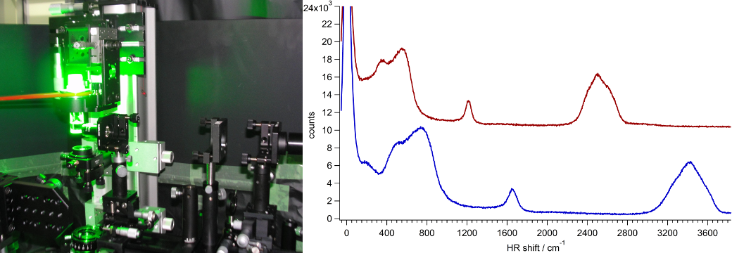
Water (blue) and D2O Hyper-Raman spectra recorded within 10 min at 532 nm excitation (400 mW)
One of the interesting research projects is to study the water structure in different aqueous systems. The advantage of the Hyper-Raman spectroscopy is that it can detect inter-molecular bands of water with high signal-to-noise ratio.
This apparatus is used for both microscopic and macroscopic Raman measurements. Currently this system has a 632.8 nm He-Ne laser and a 785 nm diode laser for excitation in a single apparatus. This gives great advantage of detecting Raman signal with wide range of excitation wavelengths. Different excitation wavelengths allow us to choose a resonant wavelength for specific sample and obtain Resonance Raman spectra, simultaneously reducing auto-fluorescence. The system combines a customised Zeiss microscope for sample placement and a peltier cooled multi-channel photon detector. Using the piezo-electric nano-positioning stage and the confocal system, near-diffraction-limit high spatial resolution Raman imaging is available suitable for biological samples (lateral resolution is sub µm while axial resolution is few µms). For macroscopic samples, a custom made rotating sample cell can be used for measurement.
Reference: Chen P. H., Shimada R., Yabumoto S., Okajima H., Ando M., Chang C. T., Lee L. T., Wong Y. K., Chiou A. & Hamaguchi H. , Scientific Reports, 6, Article number: 20097 (2016)